

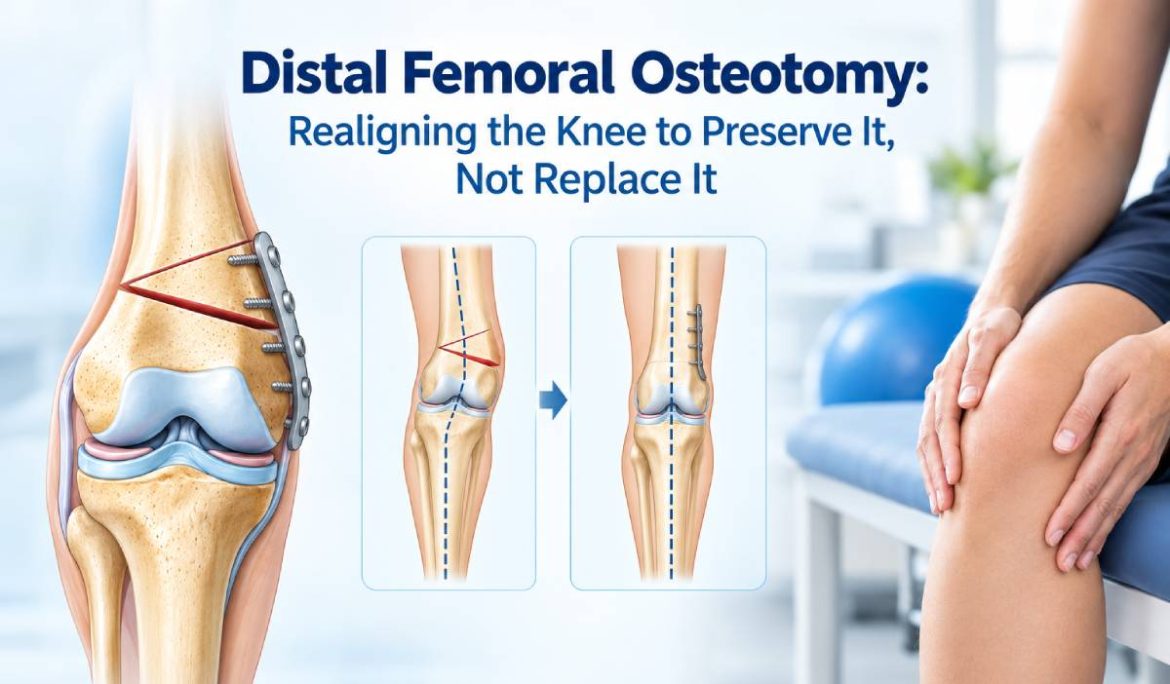
Distal Femoral Osteotomy: Realigning the Knee to Preserve It, Not Replace It
A 34-year-old homemaker from Mumbai once came to our clinic walking with her knees turned slightly inward, almost touching at every step. She had outer-knee pain for two years and had already been told by two different doctors that she would “eventually need a knee replacement.”
She didn’t need one. Not yet, and possibly not for another twenty years. What she needed was a distal femoral osteotomy – a procedure that corrects the alignment of the leg so that body weight stops overloading one side of the knee. Her own joint, her own cartilage, simply redirected to carry weight the way it was meant to.
This is the idea behind joint preservation surgery: before considering artificial joints, can we correct the underlying mechanics and explore alternatives to knee replacement that may help the patient’s natural knee last longer? Patients exploring joint-preserving options can also learn about treatments that may help save the knee from arthritis and understand how treatment choices depend on the extent of joint damage. For younger, active patients in Thane and Mumbai with a valgus knee deformity, distal femoral osteotomy may offer a joint-preserving option that many patients do not know exists.
What Is a Distal Femoral Osteotomy?
A distal femoral osteotomy (DFO) is a type of knock knee correction surgery in which the lower end of the thighbone (femur) is carefully cut and repositioned to correct the angle of the leg. The bone is then secured in its corrected position with a plate and screws while it heals, providing stability after knee osteotomy surgery.
Think of your leg’s weight-bearing line as a straight rope running from your hip to your ankle. In a healthy knee, this line passes through the centre of the joint, so weight is shared evenly. In a valgus knee – commonly called a “knock-knee” – this line shifts outward, overloading the outer (lateral) part of the knee joint.
Over years, that uneven loading wears down cartilage faster on that side, leading to early, one-sided arthritis. By cutting and re-angling the femur, a distal femoral osteotomy shifts this weight-bearing line back towards the centre, taking pressure off the damaged outer compartment and shifting it to the healthier inner part of the knee.
Distal Femoral Osteotomy vs High Tibial Osteotomy: Varus and Valgus Knees
Patients often ask why some people get surgery on the thighbone and others on the shinbone for what sounds like a similar problem. It comes down to which way the knee is bent, and where the arthritis has started.
| Feature | Valgus Knee (Knock-Knee) | Varus Knee (Bow-Leg) |
| Deformity direction | Knees angle inward, feet apart | Knees angle outward, bow-shaped legs |
| Side of knee overloaded | Outer (lateral) compartment | Inner (medial) compartment |
| Correction procedure | Distal Femoral Osteotomy (DFO) | High Tibial Osteotomy (HTO) |
| Bone corrected | Lower end of the femur (thighbone) | Upper end of the tibia (shinbone) |
| Common in | Often seen after old fractures, ligament laxity, or lateral compartment arthritis | More common overall; linked to medial compartment osteoarthritis |
Both are limb mal-alignment correction procedures, and both aim for the same outcome: redistributing weight across the knee joint so your own cartilage lasts longer.
Causes and Risk Factors for a Valgus Knee
- Previous fracture around the knee that healed in a slightly angled position
- Ligament injury, particularly on the inner side of the knee, causing gradual outward drift over the years
- Lateral meniscus tears or earlier meniscus surgery that altered joint mechanics
- Early lateral compartment arthritis, which can itself worsen the valgus angle over time
- Inflammatory arthritis (such as rheumatoid arthritis), which more frequently affects the outer knee compartment
- Growth-related or developmental knock-knee that persisted from childhood or adolescence
- Obesity and sedentary lifestyle, which accelerate wear once mal-alignment is present
In our practice across Thane and Mumbai, we commonly assess patients who need valgus knee deformity treatment after an old sports injury or fracture, as well as those whose persistent knee pain was initially dismissed as routine joint pain.
Symptoms and Warning Signs
- Pain specifically on the outer side of the knee, often worse with walking, squatting, or climbing stairs
- Visible knock-knee appearance, with knees touching or angled inward while standing
- A feeling of the knee “giving way” on uneven ground
- Swelling after long periods of standing or walking
- Gradually worsening pain over months to years, rather than from a single injury
- Difficulty sitting cross-legged or in positions requiring full knee bend, common in Indian households
- Early morning stiffness in the knee that eases with gentle movement
If outer-knee pain and a knock-kneed stance have been building up over time, seeking knock knee treatment and getting your limb alignment assessed can help identify the underlying problem.
How Is a Valgus Knee Diagnosed?
- Clinical examination – assessing the visible alignment of the legs, knee stability, and range of motion.
- Standing (weight-bearing) X-rays – taken with the patient standing, since alignment looks different lying down; this shows how much the weight-bearing line has shifted.
- Long-leg alignment X-ray – a full-length X-ray from hip to ankle, used to precisely calculate the angle of correction needed before surgery.
- MRI – to assess the condition of the cartilage, meniscus, and ligaments, and confirm how much of the joint is still healthy enough to preserve.
- Gait assessment – observing how you walk, since this affects surgical planning and expected outcomes.
This detailed assessment helps determine the most appropriate valgus knee deformity treatment, with the correction angle calculated individually to support a personalized joint preservation plan.
Treatment Options
Non-surgical Knock Knee Treatment (Early Stages)
- Weight management to reduce load on the affected compartment
- Activity modification – avoiding high-impact activities that worsen outer-knee pain
- Physiotherapy to strengthen muscles supporting the knee
- Unloader knee braces, which shift some weight away from the damaged compartment
- Anti-inflammatory medication or occasional injections for symptom control
Surgical treatment: Distal Femoral Osteotomy
When pain and deformity progress despite conservative treatment, knock knee correction surgery such as a distal femoral osteotomy may be considered for younger, active patients who are not ideal candidates for total knee replacement.
The procedure involves:
- Precise pre-surgical planning using long-leg X-rays to calculate the correction angle required for knee osteotomy surgery.
- A controlled cut in the lower femur, opened or closed to the planned angle
- Fixation with a plate and screws to hold the new alignment while the bone heals
- In select cases, combining the osteotomy with cartilage or meniscus procedures if needed
The goal is to preserve the natural joint by correcting the mechanical problem that causes uneven wear. For carefully selected patients, osteotomy may be one of the alternatives to knee replacement worth discussing with an orthopaedic surgeon.
Exercises and Lifestyle Advice
Both before and after a distal femoral osteotomy.
certain habits support better outcomes:
- Quadriceps and hamstring strengthening exercises, guided by a physiotherapist, to support the knee joint
- Low-impact cardio such as swimming or stationary cycling once cleared by your surgeon
- Maintaining a healthy body weight to reduce ongoing joint stress
- Avoiding prolonged squatting or floor-sitting, common in Indian homes, until the knee is fully assessed
- Using a walking aid temporarily if advised, to protect the joint during early recovery
- Gradual return to daily activities as guided, rather than rushing back to long work hours or household chores
Recovery Timeline
| Phase | What to Expect |
| Week 1–2 | Hospital stay of a few days; pain management; partial weight-bearing with crutches or a walker |
| Week 2–6 | Gradual increase in weight-bearing as advised; physiotherapy begins for gentle range-of-motion exercises |
| Week 6–12 | Bone healing progresses; walking without support typically begins, guided by X-ray follow-up |
| Month 3–6 | Strengthening exercises intensify; most patients return to desk work much earlier, and to daily household activity by this stage |
| Month 6–12 | Return to higher-impact activities and sport, once bone healing is confirmed and strength has been rebuilt |
Recovery is gradual by design – the bone needs time to heal fully in its new position. Rushing this stage is one of the most common causes of complications, so following your surgeon’s weight-bearing instructions closely really does matter.
Prevention Tips
- Get any old knee injury or fracture properly evaluated, even years later, if you’ve noticed a change in your leg alignment
- Don’t ignore one-sided knee pain that’s isolated to the outer or inner part of the joint – it’s often a mechanical clue, not just general wear
- Maintain a healthy body weight to reduce the speed of cartilage wear once mal-alignment is present
- Strengthen the muscles around the knee through regular, moderate exercise
- Seek an orthopaedic opinion early if you notice a visible knock-knee or bow-leg appearance developing in adulthood
When Should You Consult an Orthopaedic Surgeon?
- You have outer-knee pain along with a visible knock-knee stance and need an assessment to determine the appropriate knock knee treatment.
- Knee pain is limited to one side and has been worsening over months
- You’re under 50–55, active, and have been told your only option is a knee replacement; an orthopaedic surgeon in Thane may help you explore whether joint preservation is appropriate.
- Knee pain affects your ability to work, manage your household, or stay active in sport
- You have a history of knee injury or fracture, and your leg alignment looks different than before
- Conservative treatment such as physiotherapy and bracing hasn’t improved your symptoms after a reasonable trial
A thorough limb alignment assessment can clarify whether valgus knee deformity treatment through joint preservation is appropriate or whether another option makes more sense. The decision should be based on clinical examination and appropriate imaging.
Frequently Asked Questions
1. What is a distal femoral osteotomy used for?
A distal femoral osteotomy is used to correct a valgus knee deformity by realigning the lower thighbone. This shifts weight away from the damaged outer knee compartment and may help delay or avoid knee replacement in suitable patients.
2. Is distal femoral osteotomy the same as a knee replacement?
No. A distal femoral osteotomy preserves your own knee joint by correcting limb alignment, while a knee replacement removes and replaces the damaged joint surfaces with an artificial implant.
3. Who is a good candidate for this surgery?
Younger, active patients with early to moderate arthritis limited to one side of the knee, caused by a valgus alignment, are typically the best candidates, rather than patients with advanced arthritis across the whole joint.
4. How long does recovery take after distal femoral osteotomy?
Most patients begin walking with support within 2 to 6 weeks, return to daily activities by 3 to 6 months, and resume higher-impact activity or sport around 6 to 12 months, once bone healing is confirmed.
5. Can a distal femoral osteotomy delay the need for a knee replacement?
Yes. By correcting the underlying alignment problem, this procedure can delay the need for knee replacement by many years, and in some patients, avoid it altogether.
6. Is knock-knee always a sign I need surgery?
No. Many people have mild knock-knee without significant pain or arthritis. Knock knee correction surgery is considered when the alignment problem causes persistent pain, one-sided joint wear, or functional limitations despite conservative treatment.
7. What’s the difference between distal femoral osteotomy and high tibial osteotomy?
Distal femoral osteotomy corrects alignment at the lower thighbone and treats valgus (knock-knee) deformities. In contrast, high tibial osteotomy corrects alignment at the upper shinbone and treats varus (bow-leg) deformities.
Book a Consultation
If you’ve been told knee replacement is your only option, but you’re young, active, and your arthritis seems limited to one side of the knee, it’s worth exploring whether joint preservation is right for you. At Tushar Nursing Home, an orthopaedic surgeon in Thane can assess limb alignment and discuss distal femoral osteotomy and related joint preservation options for suitable patients across Thane and Mumbai, supported by appropriate pre-surgical imaging and planning.
Call or WhatsApp us to book an appointment, or visit our clinic for a walk-in consultation. Expertise. Precision. Mobility.
References
- American Academy of Orthopaedic Surgeons (AAOS) – OrthoInfo: Osteotomy of the Knee, orthoinfo.aaos.org
- International Society of Arthroscopy, Knee Surgery and Orthopaedic Sports Medicine (ISAKOS) – Joint preservation and osteotomy resources, isakos.com
- National Health Service (NHS), UK – Knee osteotomy overview, nhs.uk
- Indian Orthopaedic Association – Patient resources, ioaindia.org
- Journal of Clinical Orthopaedics and Trauma – Peer-reviewed literature on osteotomy outcomes in Indian patients
This article is for general patient education and does not replace an in-person medical consultation. Please consult an orthopaedic surgeon for diagnosis and treatment specific to your condition.