Distal Femur Osteotomy
What is a Distal Femur Osteotomy?
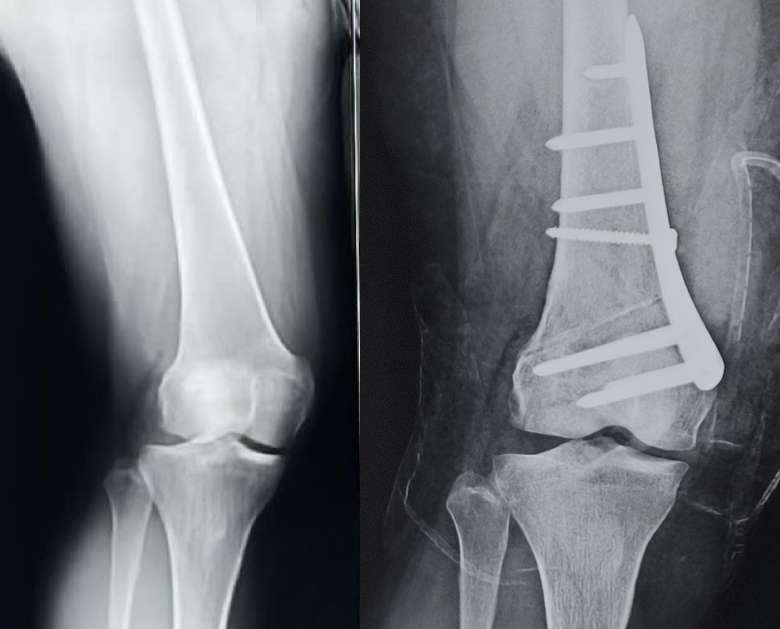
A distal femur osteotomy is a surgical procedure that involves cutting and reshaping the lower part of the thigh bone (femur) just above the knee joint. It is performed to correct knee deformities or abnormal alignment where the deformity is arising from the lower end of the femur. This surgery may be done as a stand-alone procedure or combined with arthroscopic (key hole) surgeries of the meniscus or the cartilage as per the need of the patient. Occasionally patients with severe deformities may require combined distal femur and proximal tibial osteotomy (Double osteotomy) to better distribute the correction needed.

Why is it Done?
A distal femur osteotomy may be recommended to:
- Correct imbalance due to abnormal alignment and prevent overloading of one compartment of the knee
- Correct severe bowleg (valgus) or knock-knee (varus) deformities where the deformity originates from the lower end of the femur
- Help slow the progression of knee arthritis which is secondary to abnormal loading
- Realign the knee joint in cases of malunion after fracture healing
What patient can expect before surgery?
- We have detailed clinical examination to recognize the deformity of the lower limb and the presence of additional pathologies if any.
- X-ray of the knee to evaluate the condition of the bone and the joint and ascertain the extent of the degeneration if any
- A standing scannogram is a special X-ray done to evaluate the alignment of the lower limb. The surgery aims to correct any existing deformity that is contributing to the symptoms
- MRI of the knee may be required in some patients whose complaints suggest additional injuries to the internal structures of the knee such as the meniscus and/or the cartilage.
- Preoperative physiotherapy assessment to assess the muscle strength and range of motion and to start “Pre-hab” exercises
- Anesthesia checkup to recognize potential medical issues that can affect the peri-operative course
- Additional investigations like blood tests, chest Xray, electrocardiogram (ECG), or any other test as determined by the anesthetist/physician as being essential for surgery
The Procedure
The procedure is usually performed under spinal anesthesia that may be augmented with an epidural pump for better pain control.
- An incision is made over the distal femur above the knee. The soft tissues are gently separated to expose the site of the deformity
- The femur is carefully cut partway through (osteotomy).
- A wedge of bone may be removed or the site of osteotomy opened to create a wedge-shaped space so as to restore the morphology of the lower end of the femur. Thus, the distal femur osteotomy is either closing wedge or open wedge.
- The femur is repositioned and secured with a metal plate and screws.
- The incision is closed with stitches or staples.
- The entire procedure is carried out with the help of a special X-ray machine which allows intra operative evaluation of the limb alignment.
Recovery Process
- Patients use crutches or a walker with limited weight bearing for the initial 6 weeks
- Weight-bearing is increased gradually over 6-12 weeks
- Knee range of motion exercises are started early to prevent knee stiffness
- Physical therapy focuses on regaining range of motion and strength
- Return to full activities may take 4-6 months
Benefits
- Redistributes weight across the knee joint to reduce arthritic pain
- Corrects angular deformities for better knee alignment
- For some, may delay or avoid the need for knee replacement surgery
Risks & Considerations:
Potential risks include infection, nerve injury, non-union (failure to heal), and loss of correction over time. Careful patient selection and surgical technique are important.
With proper rehabilitation, a distal femur osteotomy can provide lasting knee pain relief, correct alignment, and improved function, especially for younger active patients.


